Achilles Tendon Diagram - Schematic Of Achilles Tendon Enthesitis A Thickened Achilles Tendon Is Download Scientific Diagram : When this tendon is put under excess strain, it can become inflamed.
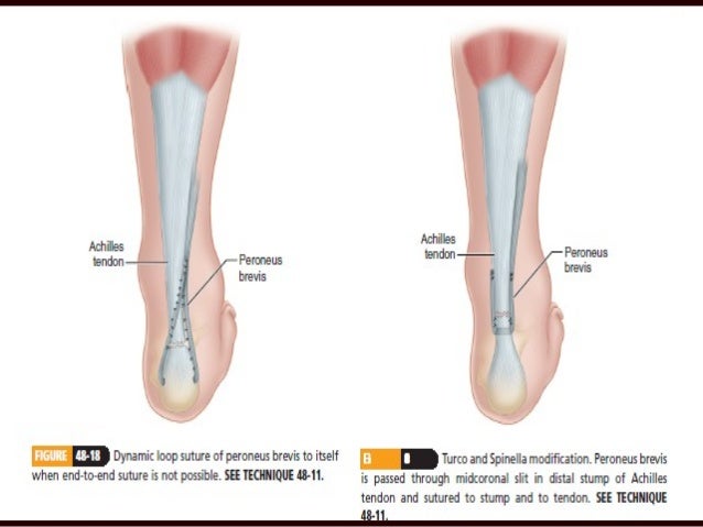
Achilles Tendon Diagram - Schematic Of Achilles Tendon Enthesitis A Thickened Achilles Tendon Is Download Scientific Diagram : When this tendon is put under excess strain, it can become inflamed.. The typical symptoms of this condition include localised achilles tendon pain that 'warms up' with activity. Your achilles tendon, which runs from your calf muscle to your heel, allows you to walk, run, jump, and flex your foot. Learn the basics of achilles tendon. Insertional achilles tendonitis involves the lower portion of the heel where the tendon inserts to the heel bone. This technique provides a description of achilles tendon reinsertion in the case of traumatic avulsion, as well as.
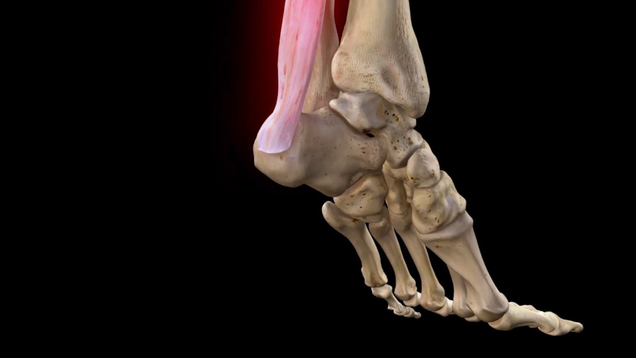
Learn vocabulary, terms and more with flashcards, games and other study tools. Also called the heel cord, the achilles tendon facilitates walking by helping to raise the heel off the ground. Ford ranger brake line diagram. It serves to attach the plantaris, gastrocnemius (calf) and soleus muscles to the calcaneus (heel) bone. Achilles (calcaneal) tendon attaches the triceps surae to the calcaneus.
Achilles Tendon Rupture Complete Anatomy Youtube from i.ytimg.com The achilles tendon is a band of tissue that connects a muscle to a bone. With aging and overuse, the achilles tendon is subject to degeneration within the substance of the tendon. Diagnosis can be made clinically. It is also the commonest tendon to rupture. Symptoms include pain, stiffness and even a bone spur. It is therefore incorrect to describe this as tendinitis. Also called the heel cord, the achilles tendon facilitates walking by helping to raise the heel off the ground. The achilles tendon or heel cord, also known as the calcaneal tendon, is a tendon at the back of the lower leg, and is the thickest in the human body.
This is in the calf, about two inches above the heel bone.
It serves to attach the plantaris, gastrocnemius (calf) and soleus muscles to the calcaneus (heel) bone. The typical symptoms of this condition include localised achilles tendon pain that 'warms up' with activity. Download this premium vector about diagram showing chronic achilles tendon tear, and discover more than 12 million professional graphic resources on freepik. This is in the calf, about two inches above the heel bone. Achilles tendon injuries can be debilitating. Insertional achilles tendonitis involves the lower portion of the heel where the tendon inserts to the heel bone. Learn the basics of achilles tendon. Achilles tendon tears or ruptures the achilles tendon is the largest tendon in the body and the achilles tendon can tear or 'rupture'. The achilles tendon joins the calf muscles to the heel bone and runs down the back of the lower leg. Achilles tendon enthesopathy is pain at the insertion of the achilles tendon at the posterosuperior aspect of the calcaneus. It is therefore incorrect to describe this as tendinitis. With aging and overuse, the achilles tendon is subject to degeneration within the substance of the tendon. The achilles tendon is the thickest in the human body.
It is named after the ancient greek. It serves to attach the plantaris, gastrocnemius (calf) and soleus muscles to the calcaneus (heel) bone. Er diagram in oracle sql developer. By allison granot, mpt, ocs, cscs, palo alto medical foundation. Achilles tendon repair, operative technique.
Midpoint And Insertional Achilles Tendonitis Insertional Achilles Tendonitis Achilles Tendonitis Achilles Tendonitis Exercises from i.pinimg.com Posted on april 3, 2019april 3, 2019. Related online courses on physioplus. Achilles tendon ruptures are common tendon injuries that occur due to sudden dorsiflexion of a plantarflexed foot, most commonly associated with sporting events. Ford ranger brake line diagram. Learn the basics of achilles tendon. Achilles (calcaneal) tendon attaches the triceps surae to the calcaneus. Insertional achilles tendonitis involves the lower portion of the heel where the tendon inserts to the heel bone. If your achilles tendon is sore or injured, you can wrap it while it heals.
This is in the calf, about two inches above the heel bone.
This is in the calf, about two inches above the heel bone. It connects the heel to the large muscles of the calf and controls the movement of the foot. The achilles tendon joins the calf muscles to the heel bone and runs down the back of the lower leg. It is named after the ancient greek. The typical symptoms of this condition include localised achilles tendon pain that 'warms up' with activity. Learn vocabulary, terms and more with flashcards, games and other study tools. Diagnosis can be made clinically. Achilles tendon ruptures are common tendon injuries that occur due to sudden dorsiflexion of a plantarflexed foot, most commonly associated with sporting events. Symptoms include pain, stiffness and even a bone spur. By allison granot, mpt, ocs, cscs, palo alto medical foundation. It is also the commonest tendon to rupture. Related online courses on physioplus. Treatment is with stretching, splinting, and heel lifts.
The achilles tendon (tendo calcaneus or tendo achillis) is the thickest and strongest tendon in the human body. Insertional achilles tendonitis involves the lower portion of the heel where the tendon inserts to the heel bone. This is in the calf, about two inches above the heel bone. This technique provides a description of achilles tendon reinsertion in the case of traumatic avulsion, as well as. It is therefore incorrect to describe this as tendinitis.
Achilles Tendon Pathology from image.slidesharecdn.com This technique provides a description of achilles tendon reinsertion in the case of traumatic avulsion, as well as. It serves to attach the plantaris, gastrocnemius (calf) and soleus muscles to the calcaneus (heel) bone. Insertional achilles tendonitis involves the lower portion of the heel where the tendon inserts to the heel bone. This is in the calf, about two inches above the heel bone. Er diagram in oracle sql developer. The achilles tendon is the thickest in the human body. With aging and overuse, the achilles tendon is subject to degeneration within the substance of the tendon. We explain what causes it, how to treat, and how to run the achilles tendon is the thickest and strongest tendon in your body, connecting your calf muscles.
Learn vocabulary, terms and more with flashcards, games and other study tools.
Insertional achilles tendonitis involves the lower portion of the heel where the tendon inserts to the heel bone. We explain what causes it, how to treat, and how to run the achilles tendon is the thickest and strongest tendon in your body, connecting your calf muscles. Er diagram in oracle sql developer. Start studying calcaneal muscle/achilles tendon. Treatment is with stretching, splinting, and heel lifts. Diagnosis can be made clinically. Musculoskeletal achilles, biceps, compression, hamstring, imaging, palpation, patella, shear, sports, tendinopathy, tendon, tendonitis. Symptoms include pain, stiffness and even a bone spur. The achilles tendon or heel cord, also known as the calcaneal tendon, is a tendon at the back of the lower leg, and is the thickest in the human body. The achilles tendon is the thickest in the human body. This protocol for achilles tendon repair is designed to provide the rehabilitation professional with a. It connects the heel to the large muscles of the calf and controls the movement of the foot. It serves to attach the plantaris, gastrocnemius (calf) and soleus muscles to the calcaneus (heel) bone.


0 Komentar